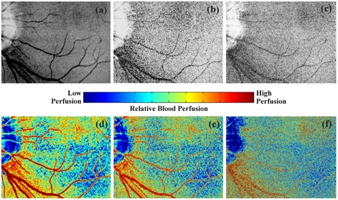
A relative measurement of blood flow changes is of major importance in the clinical domain for quantifying vascular change due to tissue injury or diseased condition. In superficial organs (viz. eye, skin), changes in blood flow are caused by different risk factors: atherosclerosis, blockage due to abnormal deposition of extracellular matrix within blood vessels, high cholesterol profile, diabetes and other inflammatory disorders like burn injury, wounds, as well as growth of benign and malignant tumours (lesions). These pathological conditions cause improper supply of oxygen and nutrients to the cells, affecting the functional and structural integrity of vasculature. In this context, Laser speckle contrast imaging (LSCI) provides a low-cost solution with high resolution probing facility among blood flow imaging modalities like: laser Doppler flowmetry, Doppler optical coherence tomography, polarization spectroscopy, photo-acoustic tomography, magnetic resonance imaging etc. This work presents speckle contrast imaging technique for in vivo imaging of microvasculature and blood perfusion in different physiological conditions. The objective is to develop computational algorithms for processing speckle images and evaluates its efficacy for quantifying changes in blood flow during abnormal state. This probing mechanism can be utilized for different applications like: detection and tracking of emboli in vascular model, changes in blood perfusion in skin flaps, changes in retinal blood perfusion due to external stimuli or various ocular pathologies, non-invasive and label free retinal angiography for the pathologies aggravating neovascularization, functional characterization of various cutaneous woundsand their stages of progression for periodic healing study etc.
- Tel: (+91) 9874375544
- Email: info@jisiasr.org


